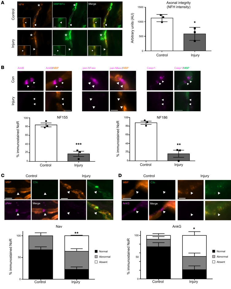
Figure 8. Extended in vivo injury selectively targeting glial membrane results in secondary axonal degeneration.
Glial mice were dosed i.p. with 50 mg/kg anti-GM1 Ab followed 16 hours later with 30 μL/g normal human serum (NHS) (injury, Inj) or NHS only (control, Con). The experiment was terminated 24 hours after NHS delivery. The site of expected nodal protein immunostaining is indicated by arrowheads. (A) At this time point there was loss of neurofilament H staining (NFH, orange) at the motor nerve terminal (MNT, asterisk) and the staining intensity was significantly reduced at the first distal node of Ranvier (NoR). (B) Normal ankyrin B (AnkB), NF155, NF186, and Caspr1 (magenta) immunostaining was assessed at distal paranodes after injury compared to control. (C) There was a further reduction in distal NoRs with normal voltage-gated sodium (Nav) channel staining (magenta) in injured Glial mice compared with control at this extended time point. (D) Additionally, the Nav channel–tethering protein AnkG was notably absent. Scale bar: 5 μm. Results are represented as the mean ± SEM. n = 3/genotype/treatment: 5–15 NoRs/mouse (median = 11, NFH intensity); 4–25 NoRs/mouse (median = 18, panNFasc); 9–23 NoRs/mouse (median = 12, Nav); and 10–28 NoRs/mouse (median = 18, AnkG) were analyzed. *P < 0.05, **P < 0.01, ***P < 0.001 by 2-tailed Student’s t test (A and B) or 2-way ANOVA with Tukey’s post hoc test (C).

