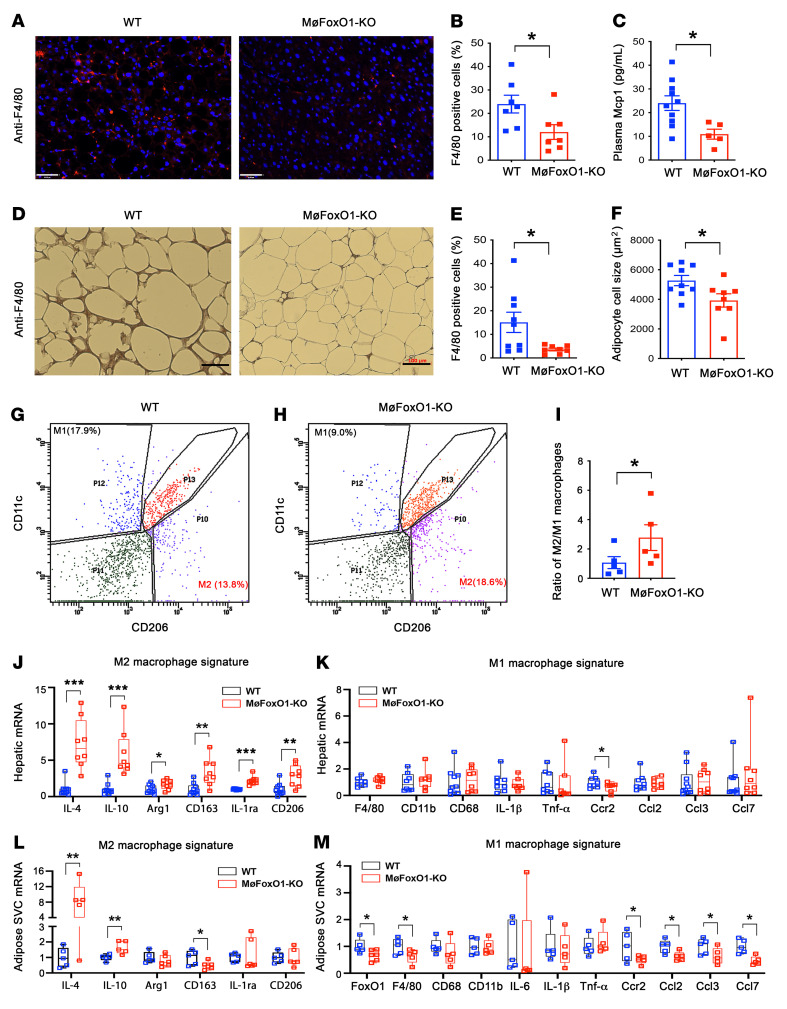
Figure 3. Myeloid FoxO1 depletion protects against HFD-elicited tissue inflammation.
MøFoxO1-KO and WT littermates (male, 8 weeks old) were fed HFD for 34 weeks. Both groups of mice were then euthanized after 16-hour fasting. Liver and epididymal fat were procured for analysis. (A) Anti-F4/80 immunohistochemistry of liver sections (original magnification, ×20). Scale bars: 50 μm. (B) Percentage of F4/80 positively stained cells in liver. (C) Plasma Mcp1 levels. (D) Anti-F4/80 immunohistochemistry in epididymal adipose tissue sections (original magnification, ×10). Scale bars: 100 μm. (E) Percentage of F4/80 positively stained cells in adipose tissue. (F) Adipocyte cell size. (G) FACS analysis of hepatic macrophages isolated from HFD-fed WT mice. (H) FACS analysis of hepatic macrophages isolated from HFD-fed MøFoxO1-KO littermates. (I) Ratio of M2/M1 macrophages in the liver. (J) Hepatic mRNA expression profile of M2 macrophage signature. (K) Hepatic mRNA expression profile of M1 macrophage signature. (L) Adipose stromal vascular cell (SVC) mRNA expression profile of M2 macrophage signature. (M) Adipose SVC mRNA expression profile of M1 macrophage signature. Data are expressed as mean ± SEM (n = 7–9). Statistical analysis in B, C, E, F, J, and L was performed using a 2-tailed, unpaired t test, and in I, K, and M using a 1-tailed, unpaired t test. *P < 0.05, **P < 0.01, ***P < 0.001.

