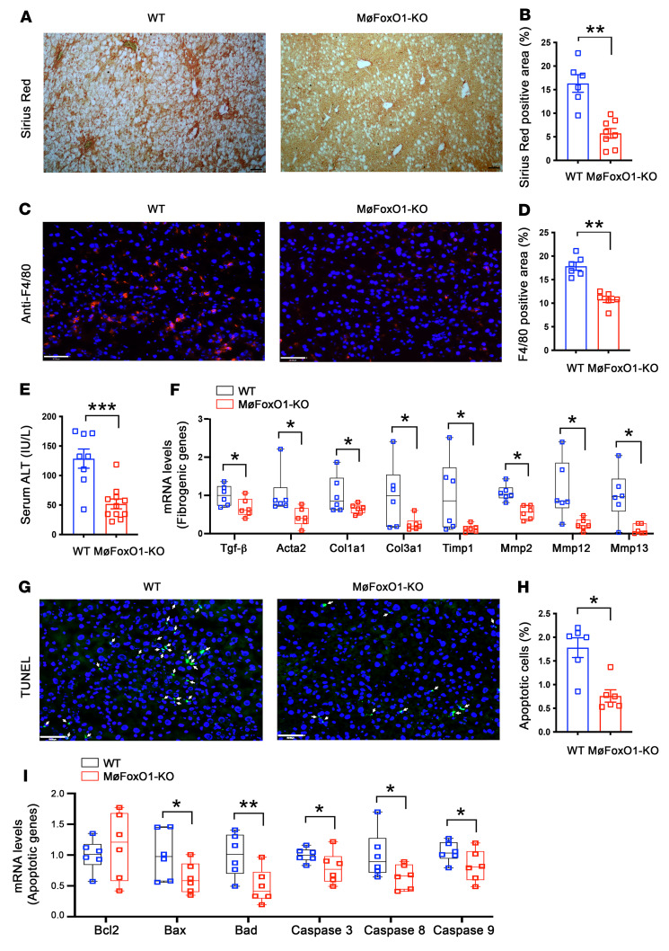
Figure 6. Myeloid FoxO1 depletion protects against diet-induced liver fibrosis.
MøFoxO1-KO and WT littermates (male, 6 weeks old) were fed a NASH diet. After 25 weeks of NASH diet feeding, mice were euthanized after 16-hour fasting. Liver tissues were subjected to histological examination. (A) Sirius red staining of liver sections (original magnification, ×10). Scale bar: 50 μm. (B) Percentage of Sirius red positively stained area of liver sections. (C) Anti-F4/80 immunostaining (original magnification, ×20). Scale bar: 50 μm. (D) Percentage of F4/80 positively stained cells in liver. (E) Serum ALT levels. (F) Hepatic mRNA levels of key genes in liver fibrosis. (G) TUNEL staining of liver sections (original magnification, ×20). TUNEL positively stained cells are marked by arrows. Scale bar: 50 μm. (H) Percentage of apoptotic cells, defined by TUNEL positively stained cells of liver sections. (I) Hepatic mRNA levels of key genes in pro- and antiapoptotic functions. Data are expressed as mean ± SEM (n = 6–11). Statistical analysis in B, D, E, and H was performed using a 2-tailed, unpaired t test, and in F and I using a 1-tailed, unpaired t test. *P < 0.05, **P < 0.01, ***P < 0.001.

