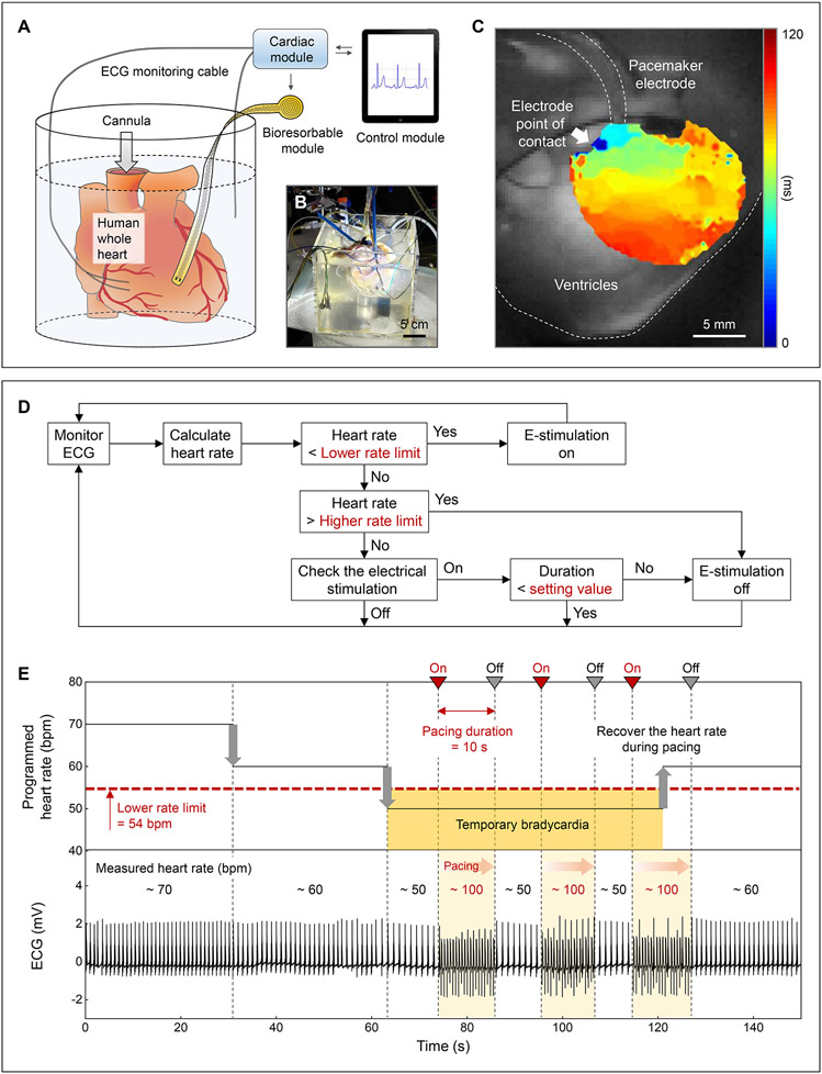
Fig. 3. Treatment of temporary bradycardia.
(A) Schematic illustration and (B) photograph of a Langendorff-perfused human whole heart model with a transient closed-loop system (dcoil = 25 mm). (C) Action potential maps obtained by optical mapping of the human epicardium. (D) Flow chart of closed-loop hysteresis pacing to activate the pacemaker upon automatic detection of bradycardia (Supplementary Text 8). (E) Programmed HR (top) and measured ECG (bottom) of a human whole heart. Set parameters: lower rate limit, 54 bpm; pacing duration, 10 s; pacing rate, 100 bpm.

