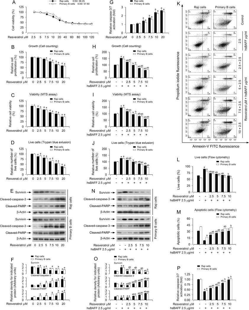
Fig. 1.
Resveratrol attenuates hsBAFF-stimulated B-cell proliferation and survival. Raji cells and primary mouse B cells were treated with resveratrol (0–120 μM) for 48 h (for IC50 and growth inhibition assay), or with resveratrol (0–20 μM) for 12 h (for Western blotting) or 48 h (for cell proliferation, viability and/or apoptosis assays), or pretreated with/without resveratrol (2.5–10 μM or 10 μM) for 1 h and then stimulated with/without hsBAFF (2.5 μg/ml) for 12 h or 48 h. (A) IC50 values and growth inhibition were determined using CCK-8 Assay Kit. (B, H) The relative proliferation was evaluated by cell counting. (C, I) The relative viability was detected by MTS assay. (D, J) The relative number of live cells was estimated by trypan blue exclusion assay. (E and N) Total cell lysates were subjected to Western blotting with indicated antibodies. The blots were probed for β-actin as a loading control. Similar results were obtained in at least five independent experiments. (F, O) The relative densities for survivin, cleaved-caspase-3, cleaved-PARP to β-actin were semi-quantified using NIH image J. (G, P) Caspase-3/7 activity was determined using Caspase-3/7 Assay Kit. (K) The percentages of live (LL), early apoptotic (LR), late apoptotic (UR) and necrotic cells (UL) were determined by FACS using annexin-V-FITC/PI staining. The results from a representative experiment are shown. (L, M) Quantitative analysis of live and apoptotic cells by FACS assay. All data were expressed as mean ± SE (n = 3 for F, L, M. O; n = 6 for A-D, G, H-J, P). Using one-way ANOVA, ap < 0.05, difference vs control group; bp < 0.05, difference vs 2.5 μg/ml hsBAFF group.

