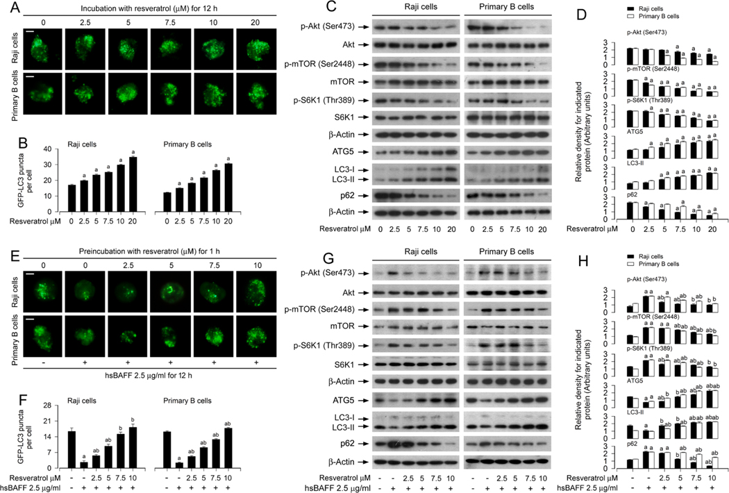
Fig. 2.
Resveratrol blocks hsBAFF-induced activation of Akt/mTOR signaling and inhibition of autophagy in B cells. Raji cells and primary mouse B cells infected with/without Ad-GFP-LC3 were treated with resveratrol (0–20 μM) for 12 h, or stimulated with/without hsBAFF (2.5 μg/ml) for 12 h following pretreatment with/ without resveratrol (2.5–10 μM) for 1 h, respectively. (A, E) Representative GFP-LC3 puncta imaging (in green) in the cells was shown by using GFP-LC3 assay. Scale bar: 2 μm. (B, F) The number of GFP-LC3 puncta per cell was quantified. (C, G) Total cell lysates were subjected to Western blotting with indicated antibodies. The blots were probed for β-actin as a loading control. Similar results were obtained in at least five independent experiments. (D, H) The relative densities for p-Akt (Ser473) to Akt, p-mTOR (Ser2448) to mTOR, p-S6K1 (Thr389) to S6K1, and ATG5, LC3-II, p62 to β-actin were semi-quantified using NIH image J. All data were expressed as mean ± SE (n = 3 for D, H; n = 6 for B, F). Using one-way ANOVA, ap < 0.05, difference vs control group; bp < 0.05, difference vs 2.5 μg/ml hsBAFF group.

