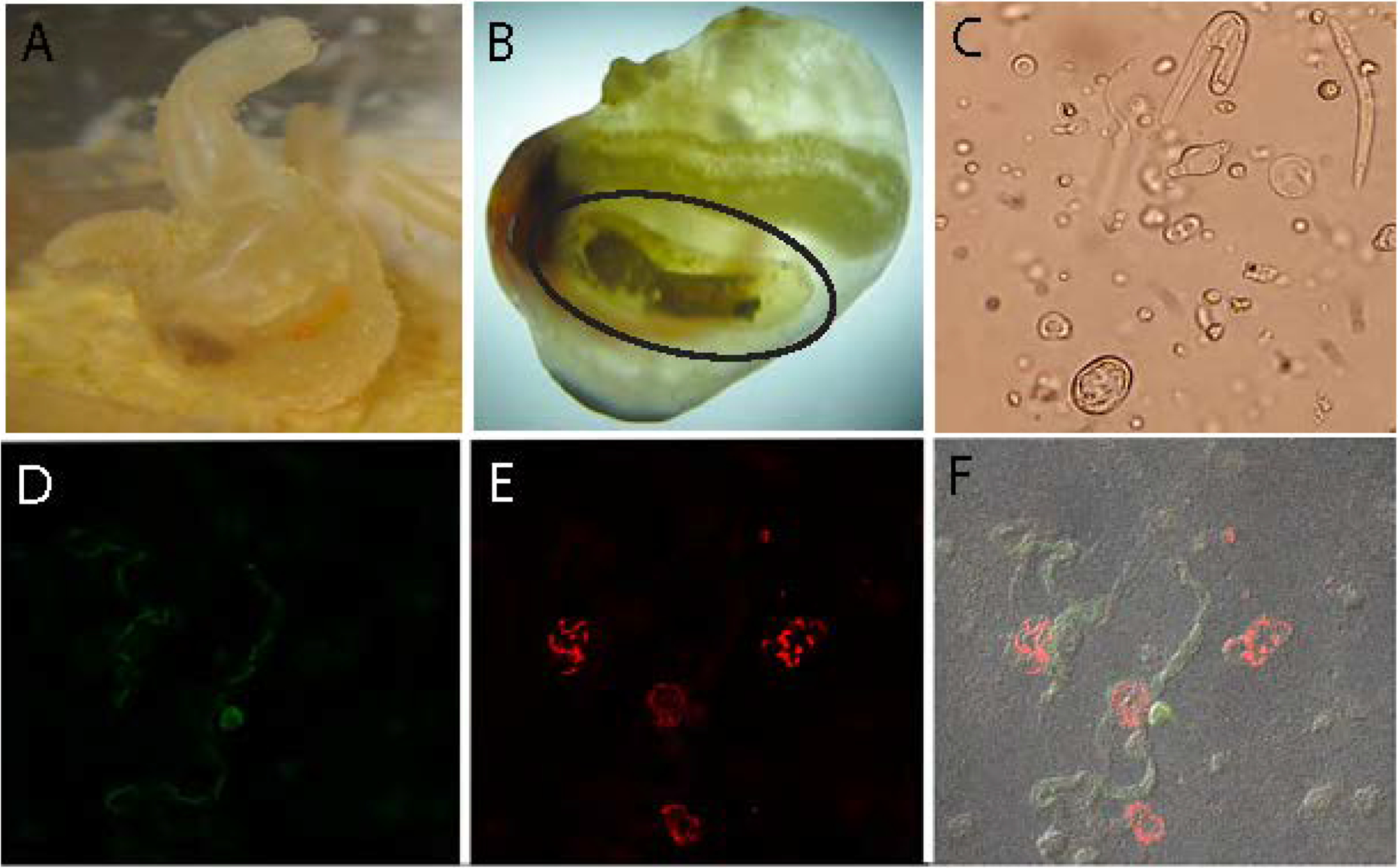
Figure 1: A multi-layered endosymbiosis between a tunicate, apicomplexan and bacterial endosymbionts.

A) Laboratory cultured Molgula manhattensis examined in this study. B) Molgula manhattensis with tunic removed to show renal sac (circled), where Nephromyces spp. spend their entire life cycle. The brown substance inside the renals sac are concretions of crystallized uric acid and calcium oxalate. C) Light microscopy photo of several Nephromyces life stages. Bacteriodes (D) and alphaprotebacterial (E) endosymbionts labeled with 16S rRNA class specific probes, using fluorescence in situ hybridization. F) The composite image of (D) and (E) showing these endosymbionts localized to different Nephromyces cells. Additional images can be seen in Figure S1.
