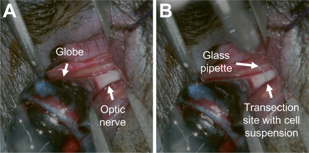
Figure 1: Transecting the optic nerve while preserving the optic nerve sheath.
(A) Picture of the surgical field demonstrating exposure of the intact optic nerve prior to transection. (B) Picture of the optic nerve following transection. A fine glass pipette pierced the optic nerve sheath at the transection site and delivered a cell suspension (turbid solution) into the space between the optic nerve ends. Please click here to view a larger version of this figure.

