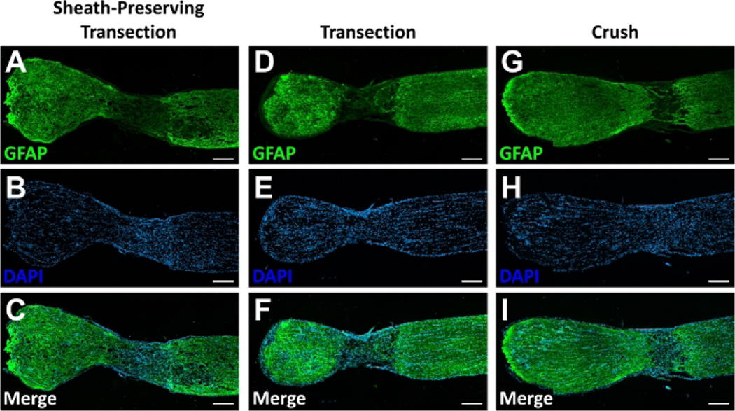
Figure 3: Sections of optic nerves following optic nerve injuries.
Representative images of longitudinal section through optic nerves 14 days after (A-C) an optic nerve sheath preserving transection, (D-F) traditional optic nerve transection, or (G-I) optic nerve crush. (A,D,G) Immunolabeling for glial fibrillary acidic protein (GFAP) delineates the extent of the lesion while (B,E,H) DNA labeling with 4′,6-diamidino-2-phenylindole (DAPI) demonstrates contiguous cellularity along the length of the optic nerve and within the lesion site. (C,F,I) Merged images demonstrating localization of GFAP-positive neural tissue and cellularity at the lesion site. Scale bars = 200 μm. Please click here to view a larger version of this figure.

