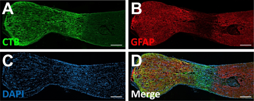
Figure 4: Section of an optic nerve following an optic nerve sheath preserving optic nerve transection.
Representative images of a longitudinal section through an optic nerve 14 days after an optic nerve sheath preserving transection with anterograde axon tracing with an intravitreal cholera toxin B subunit (CTB) injection. (A) Complete lesioning of CTB labeled RGC axons extending from the globe (left) toward the brain (right) can be observed. (B) Immunolabeling for glial fibrillary acidic protein (GFAP) delineates an extensive lesion that involves the entire diameter of the optic nerve. (C) DNA labeling with 4′,6-diamidino-2-phenylindole (DAPI) demonstrates cellularity within the lesion site. (D) A merged image demonstrating localization of CTB-labeled axons, GFAP-positive neural tissue, and cellularity at the lesion site. Scale bars = 200 μm. Please click here to view a larger version of this figure.

