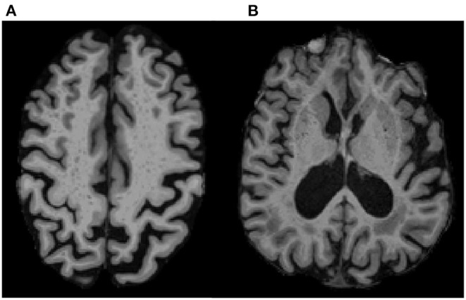
Figure 1.

ePVS Regions of Interest on T1 MPRAGE. Examples show ePVS burden in (A) the centrum semiovale (white matter; 10 mm above the lateral ventricles) and in (B) the basal ganglia (gray matter in the putamen and head of caudate in the plane of the columns of the fornix). A high intra-rater reliability was achieved for ePVS (ICC = 0.9) on a subset of 20 randomly selected participants.
