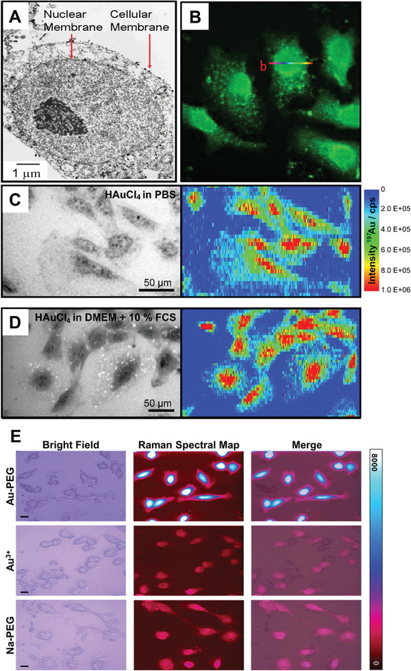
Figure 3.

Nuclear localization of GNPs formed through biomineralization. A) TEM of GNPs formed by treating HEK 293 cells with 1 mm HAuCl4 in PBS for 24 h. Reproduced with permission.[ 28a ] Copyright 2005, American Chemical Society. B) Confocal fluorescence micrograph obtained using a 488 nm excitation laser of HepG2 cells incubated in full cell media supplemented with 10 µm HAuCl4 solutions for more than 48 h. Reproduced with permission.[ 28e ] Copyright 2013, Springer Nature. Bright field optical images and 197Au+ intensity distributions from LAICP‐MS of 3T3 fibroblast cells after incubation with 1 mm of HAuCl4 for over 24 h in either C) PBS or D) full cell media. Reproduced under the terms of the Creative Commons CC‐BY license.[ 28i ] Copyright 2017, Royal Scoeity of Chemistry. E) Bright field optical images, surface enhanced Raman spectral map, and merged bright field/Raman map images of MCF7 cells treated with either 0.24 mm Au3+ admixed with 10kDa hydroxyl‐terminated PEG (Au‐PEG) or 0.24 mm Na+ with PEG (Na‐PEG) in full cell media for 4 h. Reproduced under the terms of a Creative Commons Attribution 4.0 International License.[ 28b ] Copyright 2020, Springer Nature.
