
Figure 3.
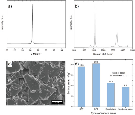
Characterization of the graphite positive electrode material: a) XRD pattern, b) Raman spectrum, c) SEM picture and d) BET surface areas of KS6L graphite. Raman data taken from previous publication.[ 46 ] SEM picture in c) shows KS6L graphite in a pristine electrode.