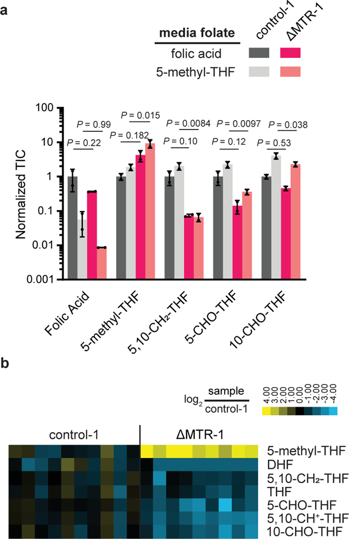
Figure 3. Loss of MTR results in the elevation of 5-methyl-THF and depletion of other folate species.
(A) Relative abundance of folate species in HCT116 control and MTR knockout cells in media with indicated folate sources. Intensities are normalized to the average of control-1 cells in folic acid (mean ± SD, n = 2, P values were determined by a one-way ANOVA comparing control to MTR knockout in the same medium followed by Dunnett’s post hoc analysis). (B) Relative abundance of folate species in individual HCT116 control and MTR knockout subcutaneous tumors (normalized to control tumor average). DHF = dihydrofolate, THF = tetrahydrofolate. TIC = total ion count.

