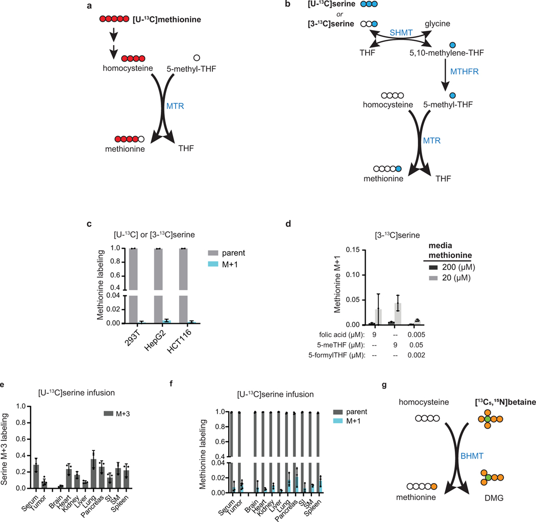
Extended Data Fig. 1. MTR is a minor source of methionine in vitro and in vivo.
(A) Schematic of methionine labeling from [U-13C]methionine. Red circles indicate 13C atoms. MTR = methionine synthase. (B) Schematic of methionine labeling from [U-13C] or [3–13C]serine. Blue circles indicate 13C atoms. MTHFR = methylenetetrahydrofolate reductase, SHMT = serine hydroxymethyltransferase. (C) Methionine labeling in cell lines after culturing for 4 h in media containing [U-13C]serine (for 293T) or [3–13C]serine (for HCT116 and HepG2) (mean ± SD, n = 2). (D) Methionine M+1 fraction from 4 h [3–13C]serine tracing in HCT116 cultured in media containing indicated methionine and folate concentrations (mean ± SD, n = 3 for each condition). Labeling of (E) serine and (F) methionine in serum, PDAC tumors, and normal tissues of male C57BL/6 mice after [U-13C]serine infusion for 2.5 h. (mean ± SD, n = 3 mice; two technical replicates were included for each tumor). (G) Schematic of methionine labeling from [13C5,15N]betaine. Orange circles indicate 13C atoms, green circles indicate 15N atoms. BHMT = betaine-homocysteine S-methyltransferase, DMG = dimethylglycine.

