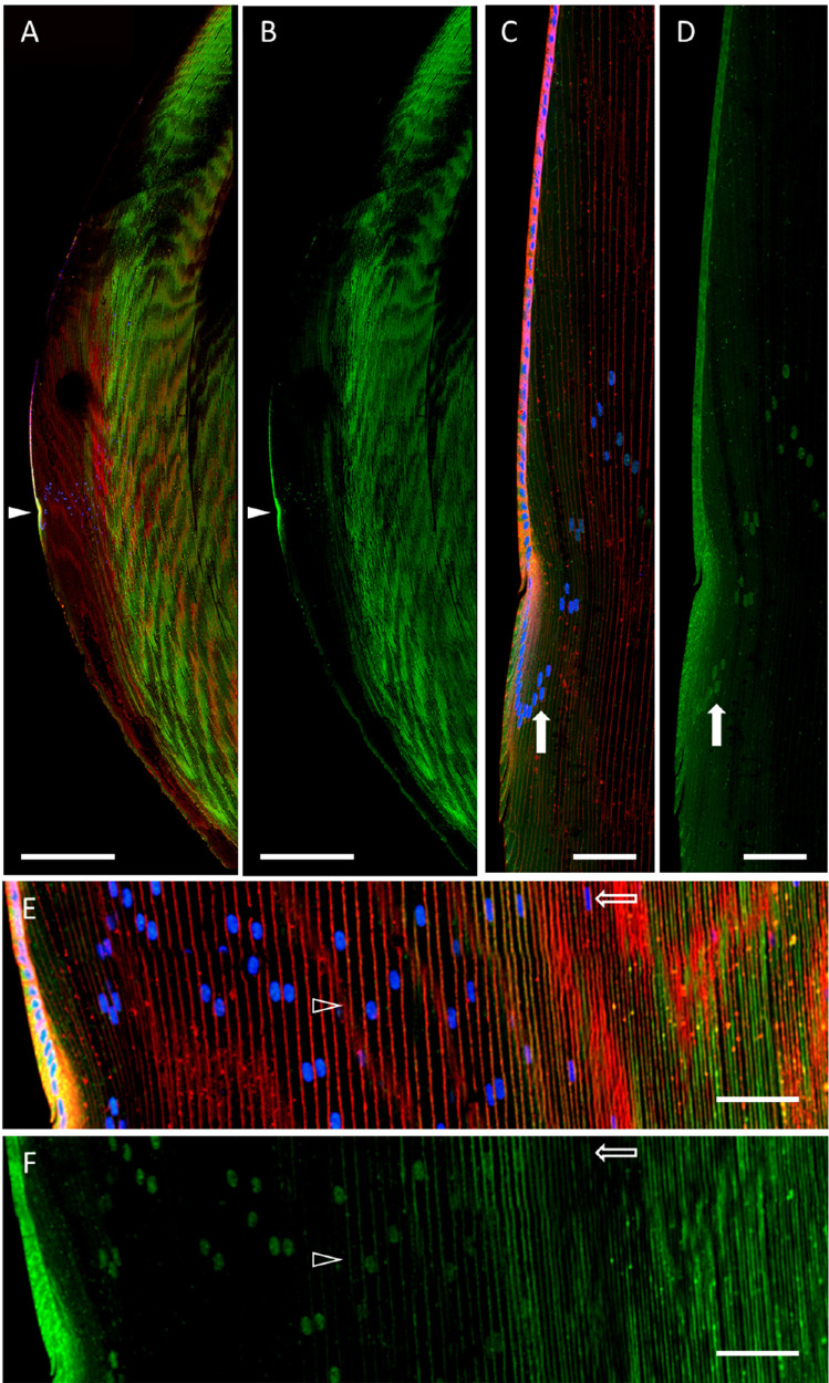
Figure 1.
AQP5 spatial expression in the bovine lens. (A) A low-magnification, high-resolution image of AQP5 immunolabeling (green) in the bovine lens with WGA labeling of plasma membranes (red) and DAPI labeling of cell nuclei (blue). The closed arrowhead denotes the lens modiolus. (B) A replicate image of A with only AQP5 immunolabeling displayed. (C) A medium-magnification, high-resolution image of the lens modiolus and nearby outer cortical fiber cells. The closed arrow indicates the nucleus of an outer cortical fiber cell. (D) A replicate image of C with only AQP5 immunolabeling displayed. The filled arrow indicates AQP5 immunolabeling in the same cell nucleus shown in B and marks the appearance of AQP5 immunolabeling in outer cortical lens fiber cell nuclei. (E) A medium-magnification, high-resolution image of the lens cortex. The open arrowhead indicates the appearance of AQP5 plasma membrane insertion. The open arrow indicates the nucleus of a differentiating cortical fiber cell. (F) A replicate image of E with only AQP5 immunolabeling displayed. Scale bars: 500 µm (A, B) and 100 µm (C–F). The open arrow indicates AQP5 immunolabeling in the same cell nucleus shown in E and marks the disappearance of AQP5 immunolabeling in inner cortical lens fiber cell nuclei.

