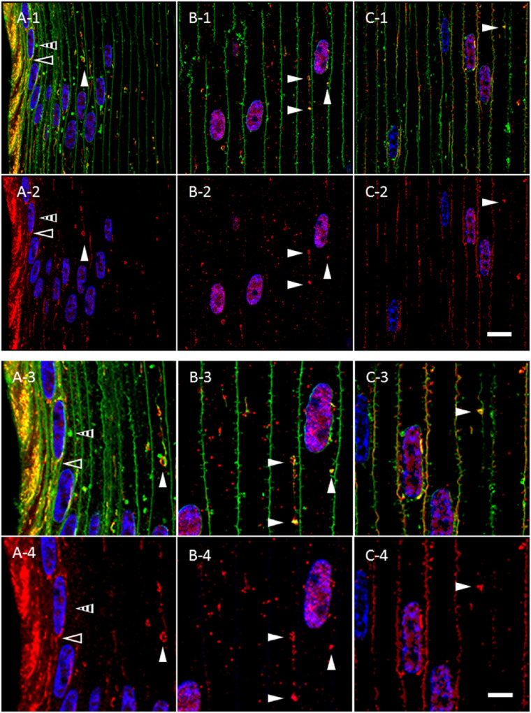
Figure 4.
AQP5-containing, cytoplasmic vesicles represent a morphologically, distinct cluster of cytoplasmic vesicles in bovine lens cortical fiber cells outside the lens modiolus. (A-1–C-1) High-magnification confocal images of AQP5 immunolabeling (red) and DiI fluorescent labeling (green) in the peripheral outer cortex (A-1), medial outer cortex (B-1), and outer cortex–inner cortex transitional region (C-1) of the bovine lens as indicated in Figure 3A. In the lens modiolus (A), linear DiI-labeled cytoplasmic compartments (open arrowheads) overlap with linear, AQP5-containing cytoplasmic vesicles. In these cells, both AQP5-negative (striped arrowheads) and AQP5-containing (closed white arrowheads) DiI-labeled cytoplasmic structures are readily observable. In the peripheral outer cortical fiber cells outside of the lens modiolus, medial outer cortex (B), and outer cortex–inner cortex transitional region (C), the spheroidal, linear DiI-labeled cytoplasmic compartments (closed white arrowheads) overlap with AQP5-containing, cytoplasmic vesicles with rare exceptions. (A-2–C-2) Replicate images of A-1 to C-1 with only AQP5 immunolabeling and DAPI labeling are displayed. (A-3–C-3) Enlarged images of DiI-labeled cytoplasmic structures are indicated by arrowheads in A-1, B-1, and C-1. Red puncta that do not colocalize with DiI represent nonspecific immunofluorescence based on normal IgG immunolabeling negative controls. (A-4–C-4) Replicate images of A-3, B-3, and C-3 with only AQP5 immunolabeling and DAPI labeling displayed. Scale bars: 10 µm (A-1, A-2, B-1, B-2, C-1, C-2) and 5 µm (A-3, A-4, B-3, B-4, C-3, C-4).

