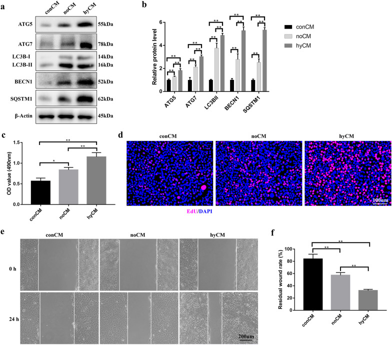
Fig. 2.
Hypoxic hBMSCs promote HaCaT cell autophagy, proliferation and migration in a paracrine manner. a Western blotting and b quantitative analysis were used to analyze the expression levels of ATG5, ATG7, LC3B-I/II, BECN1 and SQSTM1 in HaCaT cells after treatment with different conditioned media for 24 h. c MTS and d EdU assays were used to assess the proliferation of HaCaT cells after 24 h of treatment with different CM. Bar, 100. e Scratch assays and f quantitative analysis were performed to detect the migration of HaCaT cells after the cells were treated with different conditioned media for 24 h. Bar, 200 μm. Mean ± SEM. n = 3. *P < 0.05, **P < 0.01

