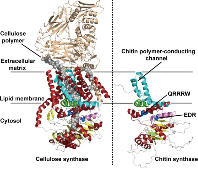
Figure 7.

Three-dimensional schematic illustration of the structure of trans-membranous chitin synthase. The left panel is the crystal structure of the bacterial cellulose synthase complex from Rhodobacter sphaeroides. Here, the cellulose polymer is shown with gray spheres. The right panel corresponds to a computed 3D structure of the C-terminal parts of chitin synthase. The crystal structure of the bacterial cellulose synthase (left panel) was used as a template for structural predictions. The highly conserved amino acid sequences include QRRRW (product binding site) and EDR (saccharide binding site), found in other glycosyltransferases such as cellulose synthases. Adapted with permission from ref (94). Copyright 2017 MDPI.
