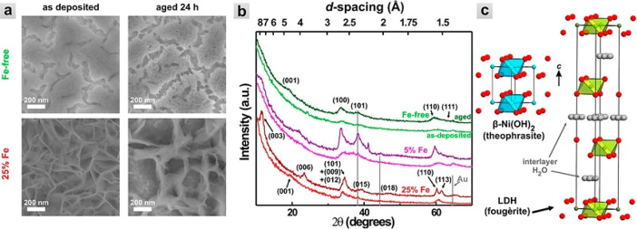
Figure 20.
(a) Scanning electron microscopy (SEM) images, (b) XRD patterns, and (c) unit cell structures for Ni(OH)2 and NiFeOxHy catalysts. (b) XRD patterns for different amounts of Fe in the NiFeOxHy catalysts. (c) Interlayer of H2O in the open LDH structure of the NiFeOxHy catalyst. Reprinted with permission from ref (169). Copyright 2014 American Chemical Society.

