
Figure 1.
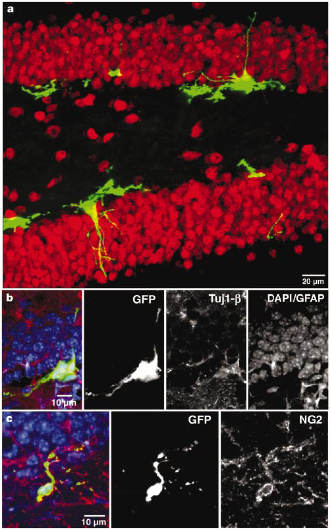
Dividing cells in the adult mouse dentate gyrus express early neural markers at 48 h after virus injection. a, Confocal micrograph (a merged image of 15 1-µm optical sections) of GFP expression in the dentate gyrus in a section that was labelled for the neuronal marker NeuN (red). No co-labelling of GFP+ cells with NeuN was observed. b, Single confocal plane of a cluster of GFP+ cells labelled with the early neuronal marker Tuj1-β (red). c, Single confocal plane of a GFP+ cell with immunoreactivity for the progenitor marker NG2 (red). In b and c, nuclei (DAPI) and GFAP are blue.