
Figure 2.
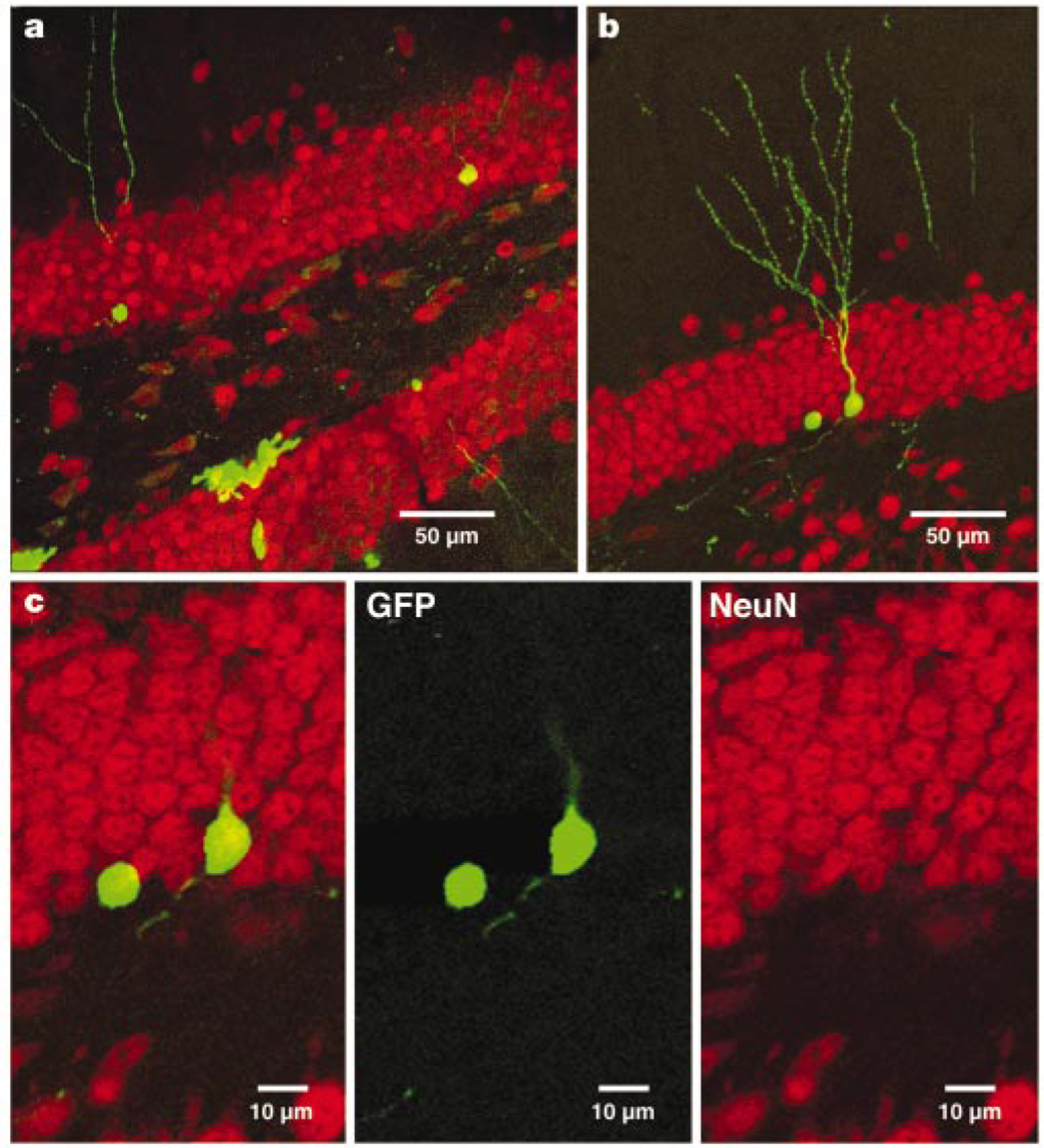
GFP+ cells in the dentate gyrus 4 weeks after virus injection express mature neuronal markers. a, Overview of GFP expression in the dentate gyrus in a section (a merged image of 27 1-µm optical sections) that was labelled for the neuronal marker NeuN (red). b, GFP+ cell (a merged image of 18 1-µm optical sections) immunoreactive for NeuN. c, Single confocal plane of the same cell, showing co-localization of GFP and NeuN.