Fig. 7.
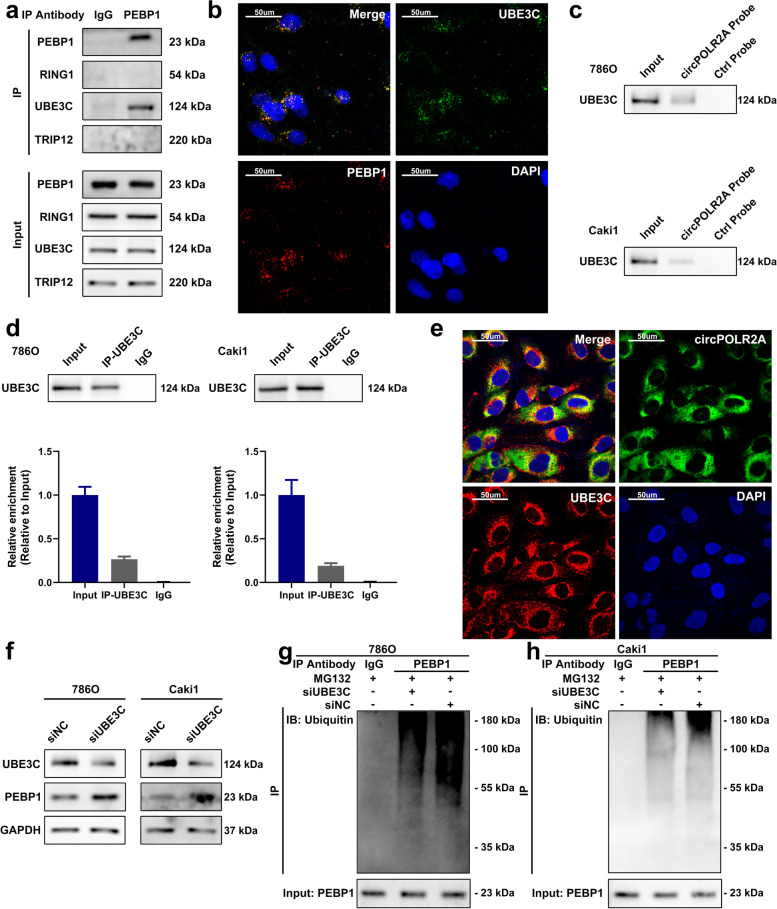
UBE3C was a ubiquitin E3 ligase which mediated the ubiquitination of PEBP1. a Co-IP with PEBP1 antibody confirmed the existence of UBE3C in the precipitates of PEBP1. b Immunofluorescence analysis of the localization of PEBP1 (red) and UBE3C (green). c RNA pull-down confirmed the binding of circPOLR2A and UBE3C. d RIP assay indicated the association between circPOLR2A and UBE3C. e RNA FISH and immunofluorescence analysis of the localization of UBE3C (red) and circPOLR2A (green). f The protein expression of PEBP1 after UBE3C knockdown in cRCC cells. Left, 786O cells; right, Caki1 cells. g, h PEBP1 ubiquitination was detected after UBE3C knockdown. 7 g, 786O cells; 7 h, Caki1 cells

