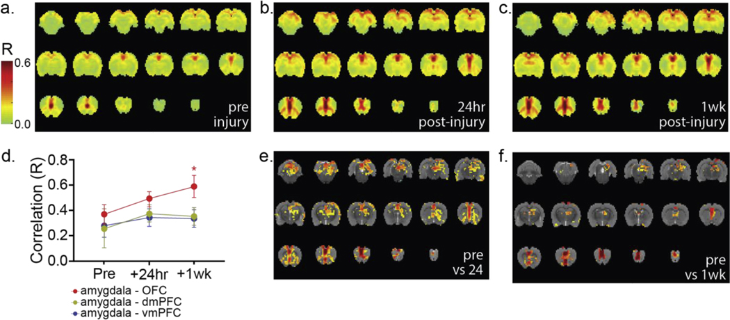
Figure 3: Localizing neurotrauma following mbTBI exposure: seed-based resting state fMRI.
We assessed rs-fMRI connectivity using the vmPFC as the seed region at (a) pre-blast, (b) post-injury day 1, and (c) 1-week post-injury in the same animals (n = 3; 6 repetitions/animal/time point). No major changes in gross network architecture were observed (a-c). Some increased connectivity was observed between network member regions, but all regions observed at pre-blast imaging remained part of the functional network at both post-injury time points. (d) Increases in region-wise (aggregate of all voxels in each region) correlated functional activity (nested 1-way ANOVA, mean±SD) between the amygdala and prefrontal cortical regions including the vmPFC (F 2,6=1.241, P=0.3541), dmPFC (F2,6=1.413, p=0.3142), and OFC (F2,6=12.09, p=0.0079) were observed across time points. OFC – amygdala correlated functional activity was significantly increased, compared to preinjury at the 1 week postinjury time point, while the remaining tracts did not differ significantly from pre-injury levels. Interestingly, at both (e) 24 hours post-injury (voxel-wise paired t-test, t=3.94 – 15.96, p=0.005 – 4.75×10⁻⁸) and (f) 1 week post-injury (voxel-wise paired t-test, t=4.46 – 11.81, p=0.005 – 5.72×10⁻⁶), rs-fMRI correlated functional activity assessed via intra-regional, voxel-wise analysis demonstrated subregional variation with significant differences at both post-injury time points in all regions of interest. In (e,f), all colored voxels have p≤0.005. Color bar at left applies to panels (a-c).

