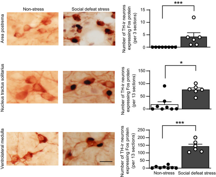
FIGURE 4.

Effects of social defeat stress on expression of c‐Fos protein in tyrosine hydroxylase‐positive cells in the area postrema, nucleus tractus solitarius and ventrolateral medulla. Photographs of sections containing cells expressing c‐Fos protein‐positive nuclear profiles and tyrosine hydroxylase‐immunoreactive (TH‐ir) cytoplasmic profiles in the area postrema, nucleus tractus solitarius and ventrolateral medulla in control mice and in mice after social defeat stress are shown (left). The number of tyrosine hydroxylase‐positive cells expressing c‐Fos protein in the area postrema, nucleus tractus solitarius and ventrolateral medulla is shown (right column). Social defeat stress increased the number of tyrosine hydroxylase‐positive cells expressing c‐Fos protein, suggesting that social defeat stress activated catecholaminergic cells in the medulla oblongata. Error bars indicate the SEM. The number of control mice and mice that received social defeat stress was 8 and 6, respectively. *P < 0.05, ***P < 0.001 vs control animals. Mann Whitney U test. Scale bar = 25 µm
