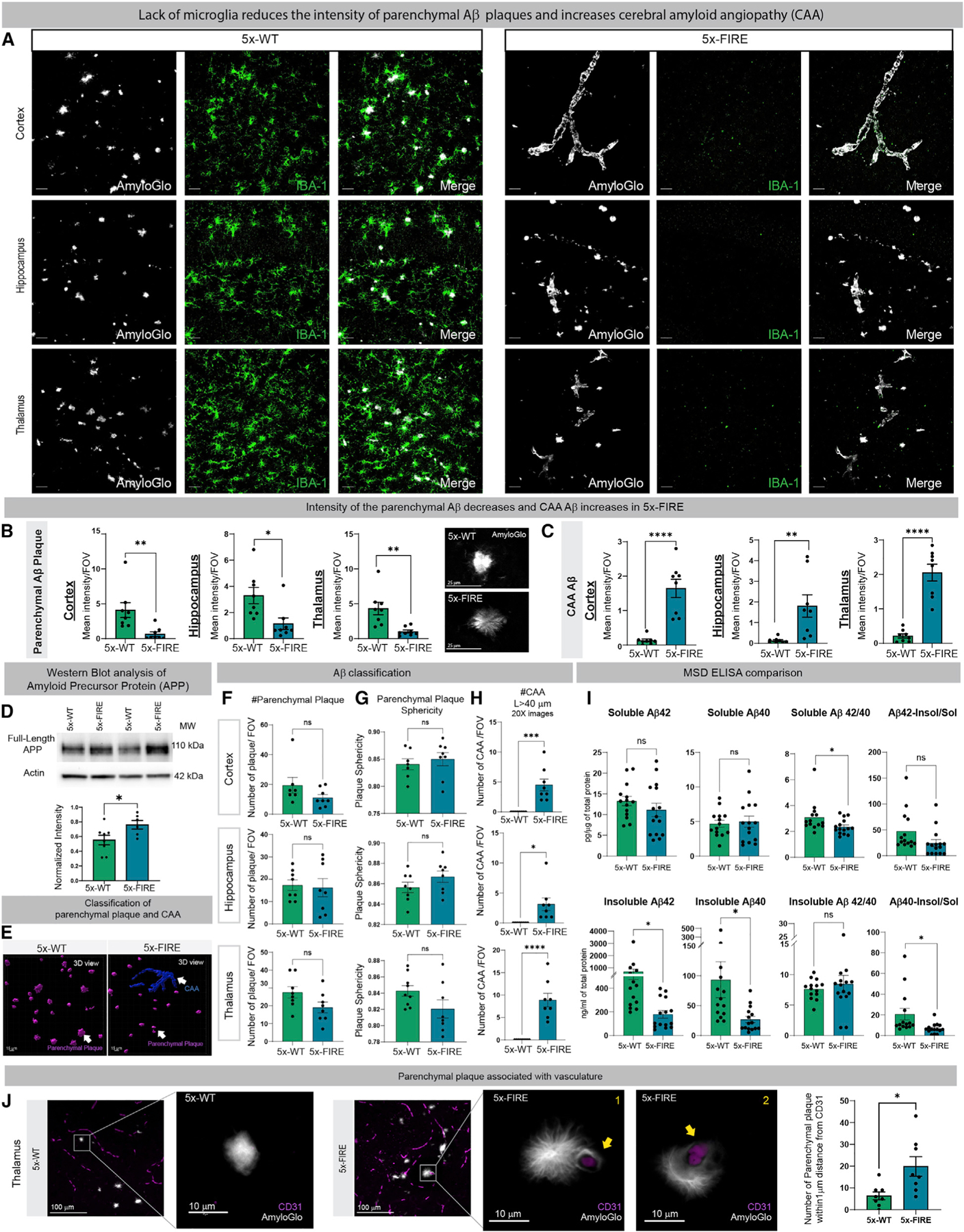
Figure 2. Microglial-deficient AD mice exhibit reduced intensity of parenchymal plaques and diminished insoluble Aβ but a robust induction of cerebral amyloid angiopathy (CAA).

(A) The 5–6-month-old 5x-WT mice exhibit parenchymal plaque deposition (Amylo-Glo, white) and clustering of IBA-1 microglia (green).
(B and C) In contrast, 5x-FIRE mice exhibit diminished plaque intensity and more diffuse morphology (B) and a robust induction of CAA (C) within all three brain regions examined.
(D) Western blots reveal a small but significant increase in human APP protein expression in 5x-FIRE mice.
(E–G) (E) Imaris image analysis was used to further classify parenchymal versus vascular amyloid pathology, revealing no significant differences in plaque number (F) or sphericity (G).
(H) However, the number of CAA deposits (H) was significantly increased in 5x-FIRE mice.
(I) ELISA analysis further reveals significantly reduced levels of insoluble Aβ40 and Aβ42.
(J) Whereas 5x-WT plaques are only occasionally observed adjacent to CD31+ blood vessels, 5x-FIRE plaques are more frequently associated with blood vessels. Scale bars, 25 µm in (A) and (B); 15 µm in (E); and 100 µm and 10 um in (J). All data presented as mean ± SEM. *p ≤ 0.05, **p ≤ 0.01, ***p ≤ 0.001, ****p ≤ 0.0001.
