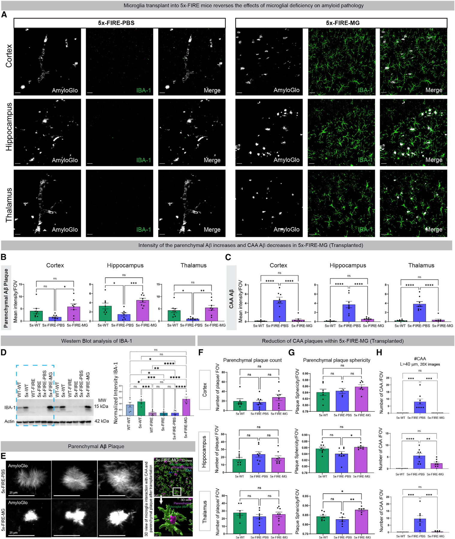
Figure 4. Adult transplantation of wild-type donor microglia prevents the effects of microglial deficiency on amyloid pathology.

(A–C) (A) Microglial repopulation leads to a reversal of the previously observed changes in amyloid distribution, increasing parenchymal Aβ intensity back to 5X-WT levels (B), while concurrently decreasing CAA (C) in comparison with PBS-injected control 5x-FIRE mice.
(D) Western blot analysis of IBA-1 further demonstrates the loss of microglia in FIRE mice and the return of IBA-1 signal following microglial transplantation.
(E) Representative high-power images of parenchymal plaques further demonstrated a shift in morphology from more diffuse filamentous plaques in 5x-FIRE-PBS mice toward more compact, intense morphology in 5x-FIRE-MG mice.
(F) Transplantation of microglia has no effect on total plaque numbers.
(G and H) In contrast, plaque sphericity within both the hippocampus and thalamus was enhanced by microglial transplantation (G) and the number of CAA deposits in all three brain regions was significantly reduced (H). Scale bars, 25 µm in (C); 25 µm, 20 µm, and 4 µm in (G). All data presented as mean ± SEM. *p ≤ 0.05, **p ≤ 0.01, ***p ≤ 0.001, ****p ≤ 0.0001.
