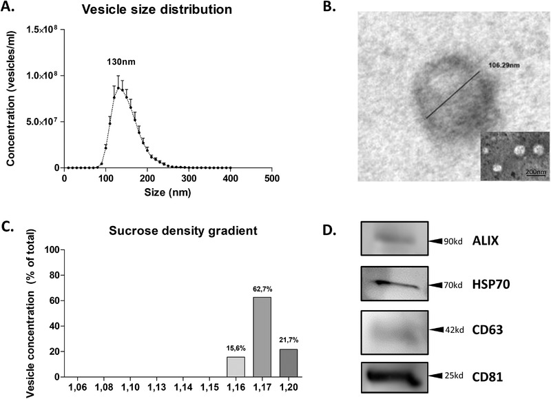
Figure 1.

Characterization of commercial milk‐derived extracellular vesicles. Within 2 h, EVs were isolated using ultracentrifugation. A) Particle size distribution of isolated vesicles was determined using a NS300. Data presented is a combination of eight separate isolations, error bars represent mean ± SEM. B) Electron microscopy confirmed spherical morphology and biolayer membrane structure. C) Sucrose density gradient following standard ultracentrifugation‐based isolation shows particles in the range of 1.16–1.20 g mL–1, which is the described range for exosome‐like vesicles. D) Western blotting confirmed the presence of EV‐markers ALIX, HSP70, CD63, and CD81.
