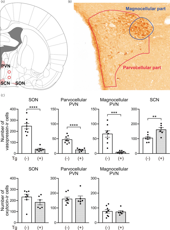
FIGURE 5.

Ablation of vasopressin neurons by intrahypothalamic injection of diphtheria toxin in transgenic rats. (A) The injection sites (red circle) are indicated on the right hemisphere according to the coronal section from the Rat Brain Atlas. 17 (B) The locations of parvocellular and magnocellular parts of the paraventricular nucleus (PVN) are drawn as lines on the photograph of vasopressin‐immunoreactive (‐IR) cells. (C) Numbers of vasopressin‐IR cells and oxytocin‐IR cells in the supraoptic nucleus (SON), the parvocellular and magnocellular parts of the PVN and the suprachiasmatic nucleus (SCN) of transgenic rats that received intrahypothalamic injection of diphtheria toxin. The numbers of vasopressin neurons on the toxin‐injected side were counted. n = 8 for control rats and n = 6 for transgenic rats. ***p < .001 and ****p < .0001 vs. wild‐type rats. Data are presented as the mean ± SEM
