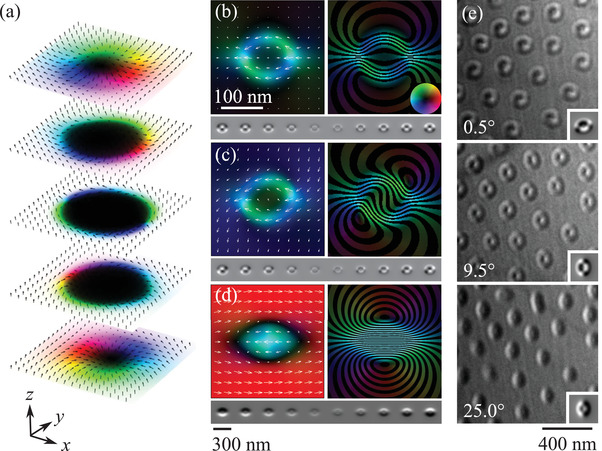
Figure 5.

a) Magnetization of a simulated type‐II bubble displayed in three dimensions as equally spaced slices. The sample surfaces lie in the xy plane and the line joining the internal domain walls is parallel to x. The uniaxial magnetocrystalline anisotropy and applied field are parallel to z. b) Projected magnetization M xy (left) and B‐field (right) for the electron beam parallel to the bubble's axis z with a defocus series for these conditions shown beneath. c) Projected magnetization and B‐field for a sample tilted 9.0° about x and 3.5° about y with respect to the electron beam. The same simulation is shown in Figure 1d. The associated defocus series is shown beneath. d) Projected magnetization and B‐field for a sample tilted 25° about y. Its defocus series is shown beneath. The simulated defocus series have the same defoci as those in Figure 3. e) Electron microscopy images of magnetic bubbles acquired with an out‐of‐plane applied field of 201 mT with defocus Δf = 0.872 mm at room temperature. Each image shows the same array of bubbles as the specimen is tilted about a horizontal axis by the angles given in the bottom left. Inserts at the bottom right show simulated images for tilt angles −9°, 0°, and 16°.
