Figure 4.
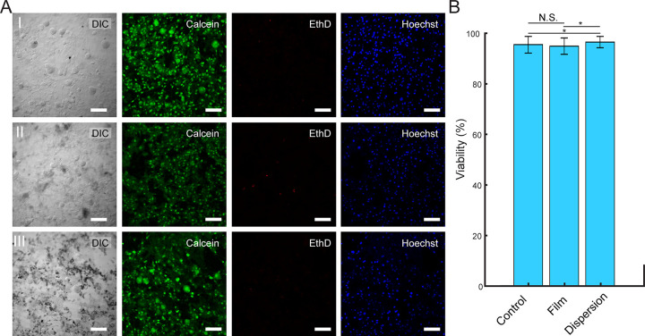
Ti3C2Tx films and dispersions do not have detectable influence on DRG neuron viability. (A) Live/Dead assay performed on DRG neurons incubated (I) without Ti3C2Tx, (II) with 25 μg/cm2 Ti3C2Tx film, and (III) with a 100 μg/mL dispersion of Ti3C2Tx flakes for 6 days. Green (Calcien AM), red (ethidium homodimer-1), and blue (Hoechst) denote live cells, dead cells, and cell nuclei, respectively. Scale bars are 100 μm. (B) Viability (%) of DRG neurons incubated without Ti3C2Tx (control), on a 25 μg/cm2 Ti3C2Tx film (film), and with a 100 μg/mL dispersion of Ti3C2Tx flakes (dispersion). Data are presented as mean ± SD (n = 5 dishes per condition, 10 images per dish). N.S. denotes no significant difference. The asterisks (*) denote statistically significant difference with p < 0.05 (one-way ANOVA and post hoc Tukey).

