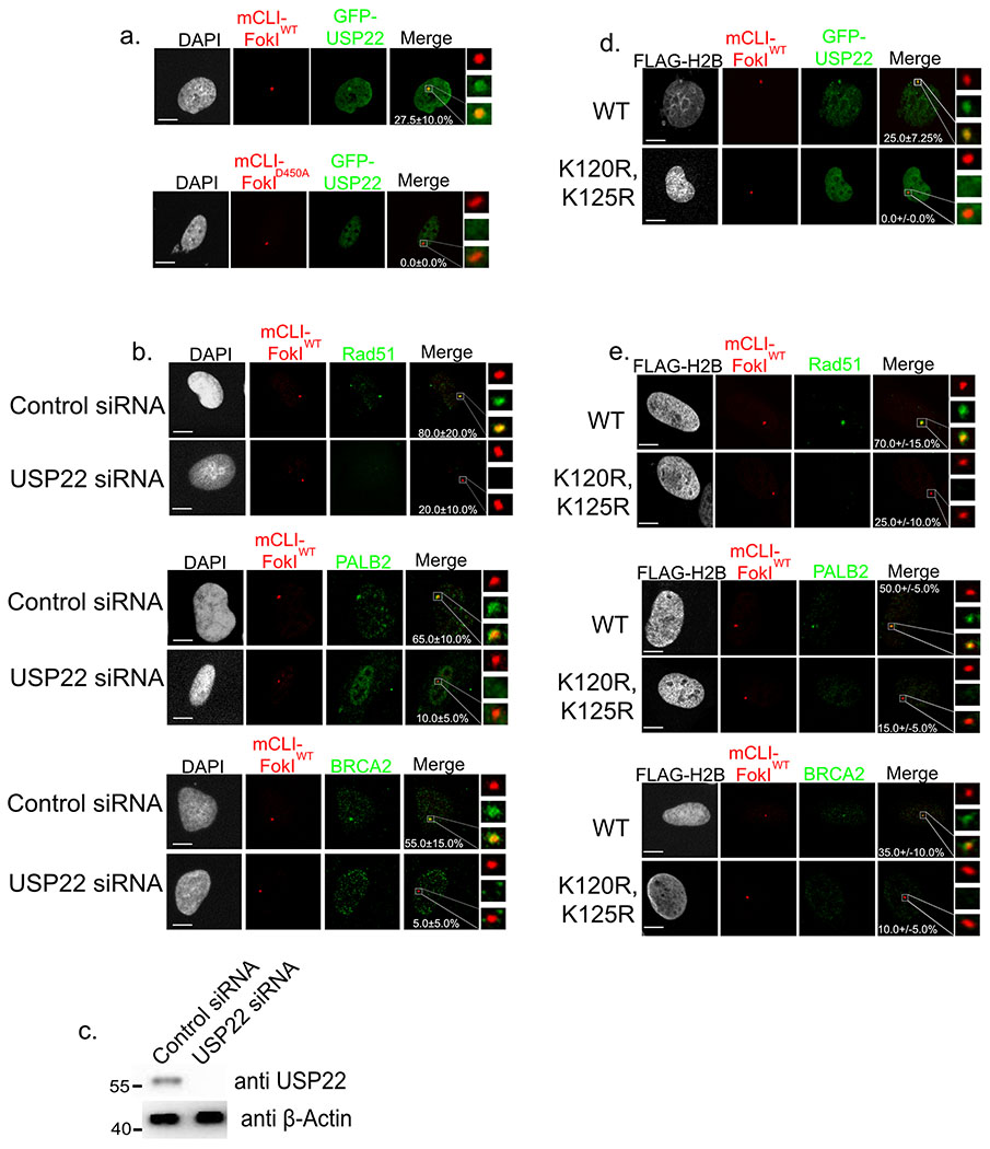
Figure 2:
Both USP22 and H2BK120ub are necessary for efficient recruitment of key HR factors to DSBs. a. U2OSFOKI cells were transfected with GFP-USP22 and treated with 4OHT and SHIELD1 ligand to induce DDR at 256X LacO array by the endonuclease mCherry-LacI-FOKIWT. Catalytically dead D450A version was used as a negative control. Percentage of cells with GFP-USP22 recruitment to array are indicated with +/− SD. White scale bars indicate 5um. 120 cells were counted for each condition for every experimental round. Experiment was done in triplicate. b. U2OSFOKI cells treated with 4OHT and SHIELD1 ligand to induce DDR at the 256X LacO array by the endonuclease mCherry-LacI-FOKIWT with/without USP22 siRNA knockdown for 48 hours. Cells were stained with antibodies against indicated proteins. Percentage of cells with recruitment of indicated protein to the array are indicated with +/−SD. White scale bars indicate 5um. 120 cells were counted for each condition for every experimental round. Experiment was done in triplicate c. Western blot against endogenous USP22 and β-Actin (loading control) after 48-hour siRNA knockdown, refers to cells from 2b. Numbers to left of western blots indicate molecular weight. d. U2OSFOKI cells were transfected with GFP-USP22 and indicated FLAG-H2B type and treated with 4OHT and SHIELD1 ligand to induce DDR at 256X LacO array by the endonuclease mCherry-LacI-FOKIWT. Percentage of cells with GFP-USP22 recruitment to array are indicated with +/− SD. White scale bars indicate 5um. 120 cells were counted for each condition for every experimental round. Experiment was done in triplicate. e. U2OSFOKI cells were transfected with indicated FLAG-H2B type for 48 hours then treated with 4OHT and SHIELD1 ligand to induce DDR at the 256X LacO array by the endonuclease mCherry-LacI-FOKIWT. Cells were stained with antibodies against indicated proteins. Percentage of cells with recruitment of indicated protein to the array are indicated with +/−SD. White scale bars indicate 5um. 120 cells were counted for each condition for every experimental round. Experiment was done in triplicate. For all statistical analysis a student’s two-tailed t-test was used to establish p-value.

