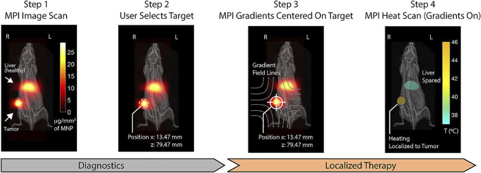
FIGURE 5.
Step 1: MPI device at 20 kHz and 20 mT produces clear visualization with SPIONs distributed in regions of pathology (tumor) and liver as the clearance organs. Step 2: The magnetic hyperthermia on the selected region is localized. Step 3: MPI gradients are shifted to center FFR solely on the target to prevent heating. Step 4: MPI gradients as heat scan at 354 kHz and 13 mT are performed on and held in the target, which could heat damage. Reproduced with permission from Tay et al., 2018a Copyright 2018, American Chemical Society. (Copyright permission shown in Supplementary Figure S1D).

