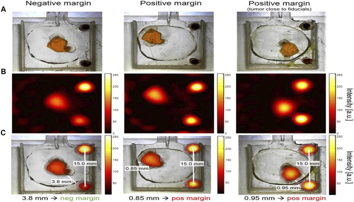
FIGURE 7.
Lumpectomy specimen phantoms, MPI signal from small-bore scanner, and co-registration images. Optical images of lumpectomy specimen phantoms. “Tumor” phantom was a space (a maximum size of ∼6.5 mm) that was filled with 0.5 mg/ml Vivotrax. The fiducials cylinders with 5.5 mg/ml Vivotrax had a 1.75 mm diameter. “Healthy tissue” was a 3D print material without SPIOs. Negative margin was considered as the distance tumor > 1 mm from the specimen’s surface; positive margin was defined as tumor≤1 mm from surface (A). MPI image was reconstructed with model-based preconditioned conjugate gradient recon (B). The image was co-registered between optical images of phantoms and MPI image, with the fiducials as controls (C). Reproduced with permission from Mason et al., 2021. Copyright 2021, Springer Nature. (Copyright permission shown in Supplementary Figure S1B).

