Figure 3.

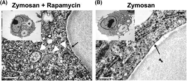
Structural comparison of rapamycin‐induced autophagosomes (A, white arrowheads) and zymosan‐induced LAPosomes (A and B, black arrow) by electron microscopy. Reprinted with permission from Springer Nature (Sanjuan et al., 2007).

Structural comparison of rapamycin‐induced autophagosomes (A, white arrowheads) and zymosan‐induced LAPosomes (A and B, black arrow) by electron microscopy. Reprinted with permission from Springer Nature (Sanjuan et al., 2007).