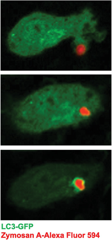
Figure 4.

Visualization of LC3‐GFP recruitment to a LAPosome containing zymosan A–Alexa Fluor 594 by confocal imaging. The still images are from time‐lapse imaging of LC3‐GFP+ macrophages’ phagocytosis of zymosan A–Alexa Fluor 594, followed by recruitment of LC3‐GFP to the zymosan‐containing LAPosome. Top, t = 10 min; middle, t = 20 min; bottom, t = 30 min.
