Figure 5.

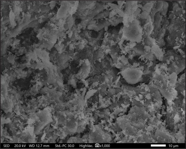
SEM micrographs of the apical third of the dentine surface of the negative control group (x1000)
SEM: Scanning electron microscope

SEM micrographs of the apical third of the dentine surface of the negative control group (x1000)
SEM: Scanning electron microscope