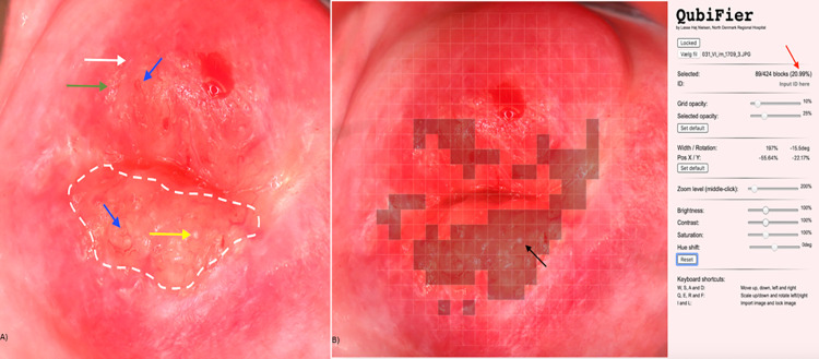
Fig 1. Digital image showing female genital schistosomiasis associated pathognomonic lesions of the cervix (picture A) and the same lesions being digitally marked by QubiFier to determine the cervical lesion proportion (picture B).
The left picture (A) shows an area, which contains homogenous yellow sandy patches appearing as yellow colored area (indicated by white dashed line). The differently colored arrows indicate the other pathognomonic lesion types: a grainy sandy patch (white arrow), clustered grainy sandy patches or rice-grain shaped sandy patches (green arrow) and a rubbery papule colored in beige with an uneven surface (yellow arrow). Abnormal blood vessel (rounded, uneven-calibered, corkscrew or convoluted) are indicated by the blue arrow. The right picture (B) shows squares of the grid marked digitally containing any types of pathognomonic lesions. The red arrow shows the proportion of the cervix covered by any type of pathognomonic lesions.

