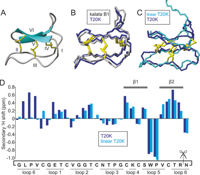
Figure 5.

Solution structure of the prototypic antiproliferative cyclotide T20K. (A) Solution structure of T20K as a ribbon diagram. β sheets are represented by blue arrows (showing direction of peptide chain), disulfide bonds in yellow, and cysteine residues numbered I-VI. (B) Backbone structure of T20K (blue) overlaid with that of kalata B1 (grey, PDB: 1nb1). (C) Backbone structure of T20K (blue) overlaid with that of linear T20K (cyan). (D) Secondary αH shift analysis of T20K (blue) and linear T20K (cyan). β strands and intercysteine loops as indicated. The amino acid sequence is written cyclic as per T20K with scissors intersecting the termini of linear T20K.
