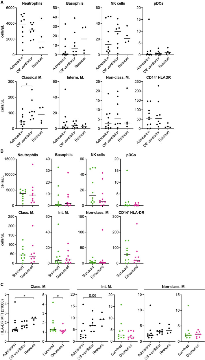
FIGURE 2.

Differences in myeloid subsets between COVID‐19 survivors and non‐survivors. A, Total cell counts for granulocytes, NK cells, plasmacytoid DCs (pDCs) and classical (Classical M.), intermediate (Interm. M), non‐classical (Non‐class. M.) and CD14+ HLADR monocytes in severe COVID‐19 survivors at different time points. B, Comparison of granulocyte and other myeloid cell subset counts at ICU admission between severe COVID‐19 survivors and non‐survivors. C, Changes in HLA‐DR mean fluorescence intensity (MFI) at different time points (left) and at ICU admission between severe COVID‐19 survivors and non‐survivors (right) in each monocyte subset. Multiple comparisons (panel A, C) were computed by Kruskal‐Wallis test with Dunn's correction. Single comparisons (panel B, C) were computed by Mann‐Whitney test. Asterisks indicate the level of significance. *P < .05
