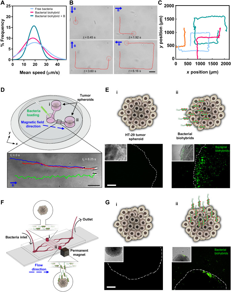
Fig. 3. Motility characterization, external magnetic control, and tumor spheroid localization of magnetic bacterial biohybrids.
(A) 2D swimming velocity analyses of free bacteria and bacterial biohybrids with or without applied external magnetic field. (B) Magnetic control of bacterial biohybrids by changing the applied field (10 mT) direction by 90° turns. B represents the magnetic field vector. Scale bar, 10 μm. (C) 2D swimming trajectories of bacterial biohybrids under external magnetic field control. (D) A magnetic guidance setup with three reservoirs, two of which contain tumor spheroids (i and ii), connected by narrow channels to the loading reservoir. A uniform magnetic field (26 mT) along the x axis was created with a permanent magnet setup. Scale bar, 10 μm. (E) Schematics and microscopy images of the tumor spheroids in reservoirs i and ii. Scale bar, 100 μm. (F) A microfluidic system with branched channels and two reservoirs (i and ii) with one tumor spheroid in each. A small permanent magnet was placed next to the reservoir ii to generate a magnetic field gradient. (G) Schematics and microscopy images of the tumor spheroids in reservoirs i and ii. Scale bar, 100 μm.

