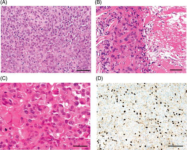
FIGURE 1.

Proximal versus distal histology. (A) Conventional‐type/distal‐type epithelioid sarcoma. The tumour is composed of sheets of essentially uniform and relatively small to medium‐sized cells with rounded to ovoid vesicular nuclei, prominent nuclei and moderate amounts of eosinophilic cytoplasm. The cells have a somewhat histiocytoid‐appearance, and cellular atypia is minimal. A mild chronic inflammatory infiltrate, largely of small lymphocytes, is intermingled (haematoxylin and eosin, ×100). Scale bar, 100 μm. (B) Conventional‐type/distal‐type epithelioid sarcoma. Nests and sheets of epithelioid cells, here with abundant eosinophilic cytoplasm, are present adjacent to a large area of fibrinoid material/incipient geographic tumour necrosis (right of field) (haematoxylin and eosin, ×200). Scale bar, 50 μm. (C) Proximal‐type epithelioid sarcoma. This tumour is composed of monotonous sheets of large, epithelioid and polygonal cells with ovoid vesicular nuclei and prominent, sometimes multiple nucleoli and abundant, palely eosinophilic cytoplasm. In many cells, the combination of extensive eosinophilic cytoplasm and eccentrically oriented nuclei give the cells marked rhabdoid appearances (haematoxylin and eosin, ×400). Scale bar, 25 μm. (D) Conventional‐type/distal‐type epithelioid sarcoma. Immunohistochemistry for INI1 (SMARCB1). This protein is absent in nuclei in approximately 90% of both classic‐type and proximal‐type epithelioid sarcoma, and here, the lesional nuclei show diffuse absence of expression of INI1. This is in contrast to the surrounding smaller numbers of lymphocytes and local stromal and endothelial cells, which show strong expression of the intact protein in their nuclei (immunoperoxidase, ×200). Scale bar, 50 μm
