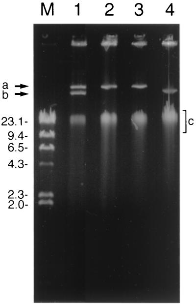
FIG. 2.
Agarose gel illustrating the plasmid profiles of A. globiformis D47 and mutants. Lane 1, strain D47; lanes 2 and 3, degrading mutants; lane 4, nondegrading mutant; lane M, markers (lambda DNA cut with HindIII), in kilobases. Positions of the two plasmids (arrows a and b) and chromosomal DNA (area c) are indicated.

