FIGURE 1.

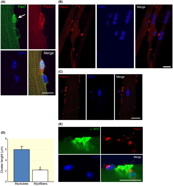
Localization of Piezo1 channels in skeletal muscle cells by confocal microscopy. A, Immunostaining for Piezo1 channels in a Pax7‐positive satellite cell (SC) (arrow) attached onto a FDB fibre (24 h post plating, 50 SCs were analysed). Note the close by Pax7‐negative nucleus likely belonging to the skeletal muscle fibre. B, Representative Piezo1 channel clusters detected in differentiating myotubes (7 day post plating). C, Immunostaining revealed Piezo1 channels organized in scattered clusters also in myofibres. Note the different scale bars in (B) and (C). D, Comparison of cluster length detected in myotubes and myofibres (n = 32 and 451 clusters analysed in myotubes and myofibres; ‡ P < .0001, Mann–Whitney test). E, Localization of Piezo1 channel clusters at a nearby endplate region of a myofibre (24 h post plating). Scale bars: 10 μm in (A) and 20 μm in (B), (C) and (E). Each group of confocal experiments were carried out on at least three independent cell culture preparations
