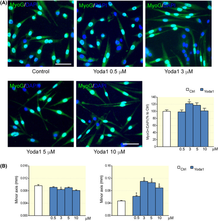
FIGURE 3.

Dose–response effect of Yoda1 on myogenic precursors. A, Representative images and graph summarizing the percentage of MyoG‐positive cells cultured in differentiation medium for 72 h in controls and in the presence of different Yoda1 concentrations (control: n = 64 optical fields; 0.5 μM: n = 38; 3 μM: n = 62; 5 μM: n = 46; 10 μM: n = 37, † P < .01, ANOVA, Dunnett's post hoc test). B, Effect of Yoda1 on cell morphology (control: n = 192 optical fields; 0.5 μM: n = 185; 3 μM: n = 293; 5 μM: n = 244; 10 μM: n = 175, * P < .05, † P < .01 and ‡ P < .001, ANOVA, Dunnett's post hoc test) after a treatment of 72 h. Experimental replicates n = 2; data from two independent experiments
