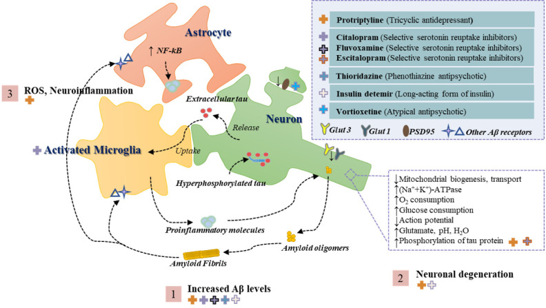
Figure 1.

Overview of the pathogenesis of AD triggered by extracellular and intravascular Aβ deposition. Step 1 shows the interaction of Aβ oligomers and fibronectin with neuronal cells through several receptors which in turn triggers an overall increase in brain Aβ levels. Step 2 shows that neurons suffer from impaired mitochondrial bioenergetics and glucose homeostasis and thus degeneration. Step 3 illustrates that the neuroinflammatory environment and oxidative stress to which the neuronal cells are exposed can exacerbate the pathological process of AD. In addition, the top right corner of the figure shows the anxiolytic drugs reported in the literature for the treatment of AD models, with the crosses on the left corresponding to the respective antipathogenic mechanism in the figure and the classification of the drug in parentheses. Abbreviations: Aβ, amyloid peptide; GLUT, glucose transporter; NF-κB, nuclear factor kappa light chain enhancer of activated B cells; ROS, reactive oxygen species; PSD95, postsynaptic density protein-95.
