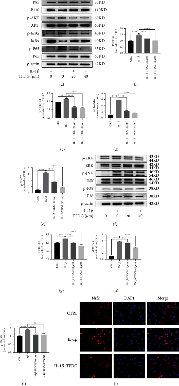
Figure 6.

TFDG inhibits inflammation-related pathways induced by IL-1β. (a–i) The protein expressions of phosphorylated PI3K, AKT, p65, IκBα, ERK, JNK, and p38 were analyzed by western blotting. The grey values normalized with control group were quantified by ImageJ. The bar graph shows the mean ± SD of data (n = 4). ∗p < .05, ∗∗p < .01, ∗∗∗p < .001, and ∗∗∗∗p < .0001. (j) P65 protein expressions were detected by immunofluorescence. Scale bar: 100 μm.
