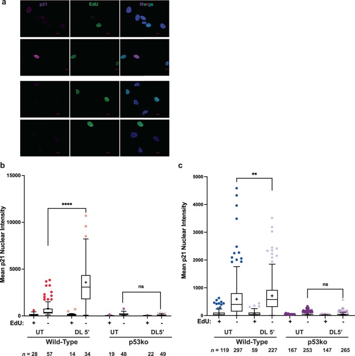
Extended Data Fig. 6. Telomeric 8oxoG increases p53-dependent p21 expression in non-replicating cells.
(a-c) 23 hours after treatment, wild-type and p53ko RPE FAP-TRF1 (b) and BJ FAP-TRF1 (c) cells were pulsed with EdU for 1 hour, and then analyzed by microscopy for p21 and EdU staining. In each condition, cells were categorized as EdU + or – populations, and the nuclear p21 signal intensity was graphed. Representative IF images are shown in panel a, scale bar = 10 μm. The number n of cells analyzed for each condition from two independent experiments is shown. Tukey box plot shows medians (bar), means (+), 25th to 75th interquartile range (IQR), and whiskers showing 25th or 7th percentile ± 1.5x the IQR. Data analyzed by two-way ANOVA (**p < 0.01, ****p < 0.0001).

