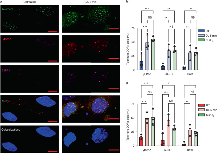
Fig. 4. Telomeric 8oxoG promotes a localized DDR.
a, Representative IF images showing γH2AX (red) and 53BP1 (purple) staining with telomeres (green) by telo-FISH for BJ FAP-TRF1 cells 24 h after no treatment or 5 min DL. Colocalizations panel shows NIS-Elements-defined intersections between 53BP1 and/or γH2AX with telomeres. Scale bars, 10 μm. b,c, Quantification of percentage of cells exhibiting telomere foci colocalized with γH2AX, 53BP1 or both for BJ (b) and for RPE (c) FAP-TRF1 cells 24 h after 5 min DL or 2.5 mM KBrO3 treatment. Error bars represent the mean ± s.d. from three independent experiments in which more than 50 nuclei were analyzed per condition for each experiment. Statistical significance determined by two-way ANOVA (ns, not significant; *P < 0.05; **P < 0.01; ***P < 0.001).

