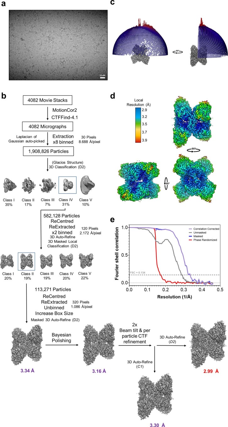
Extended Data Fig. 2. Image processing workflow of the GYS1:GYG1ΔCD inhibited state.
a, Representative K3 micrograph of the GYS1:GYG1ΔCD inhibited state from 4082 micrographs collected. b, Processing flow chart of the GYS1 + GYG1ΔCD inhibited state. c, Angular distribution of the 3.0 Å GYS1:GYG1ΔCD inhibited state map. d, Local resolution variation of the 3.0 Å GYS1:GYG1ΔCD inhibited state map. e, FSC curve of the 3.0 Å GYS1:GYG1ΔCD inhibited state map.

